|
Gefangenschaftskrankheiten beim Elefanten Captivity
Disorders in Elephants D.A. Fagan,
J.E.Oosterhuis, and A. Roocroft. |
||||||||||||
|
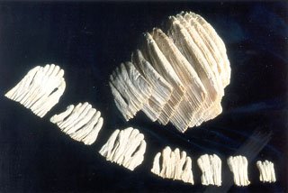
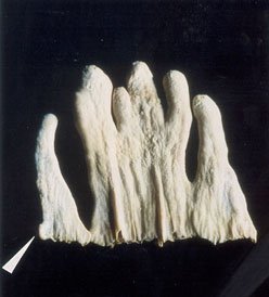
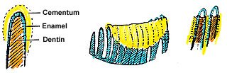
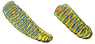
DEVELOPMENTAL PROCESSES OF MOLARS The elephant's molar dentition, perhaps surprisingly, features several unique characteristics, notably the manner in which each molar tooth is created by the fusion of a dozen or more successive dental plates ( see Figure 3). A predetermined number of these enamel plates are formed as embryonic denticles evolving from the replicating dental lamina. These still-developing plates or tooth buds are shaped like small human hands ( see Fig. 4). They are composed of tooth forming cells, which differentiate into the basic odontoblastic components of mammalian teeth, and in due course, manufacture the enamel, dentin and cementum necessary to create a single dental plate ( see Fig. 5). As this progression of individual plates matures, they fuse together to create a " package of dental plates", which then becomes the molar tooth. Many developmental dental maladies such as supernumerary teeth, gemination, and crown or root dilaceration can and do occur at this stage of development resulting in the eventual malocclusion of a deformed or nonfunctional tooth. This process has been described by many authors including Colyer, Kingdon, Mitchell and Van der Merwe. For a more detailed definition of gemination and related terms, see Ferreira et al. Briefly, however, gemination arises when two tooth buds fuse together to form a single tooth, and dilaceration refers to a marked angular curvature or distortion in the normal anatomical form of the crown or root portion of the tooth.
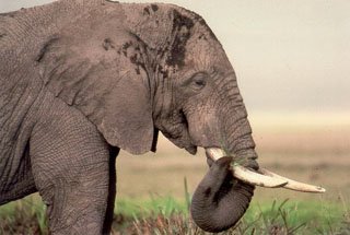
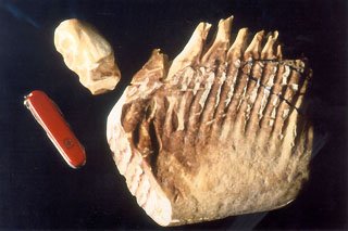
The molars, contained in the massive tuberosity of the elephant's maxilla or body of its mandible, are composed of a number of parallel plates or vertical laminae of enamel surrounded by dentin and cementum. The enamel, by virtue of its superior hardness, is less susceptible to abrasive attrition during mastication, and eventually remains standing above the more easily worn dentin and cementum, resulting in an uneven occlusal grinding surface. Each of the individual dental plates is joined throughout the body of the molar tooth, which thus creates a tooth with 15 to 30 functional apical openings (see Fig 6 ), and a "grinding surface" on the mature molar, which permits it to function with its opposing tooth much like two opposing course farrier's rasps providing the elephant with a most efficient masticatory apparatus. On the occluding or grinding surface, the pattern formed by these worn enamel plates distinguishes the Asian elephants' molars with a squashed ovoid pattern, from the African elephant with a distinctive diamond pattern (see Fig 7). In fact, it is this pattern of the molars which provides the origin of the African elephant's scientific name Loxodonta - from the Greek word loxos - meaning oblique.
The molars of the African elephant are considerably larger than those of the Indian, and are well endowed with a more substantial portion of enamel for efficient mastication of a courser and harder diet. Their first or primary set of molars, sometimes referred to as the Milk teeth, gradually give way to a second set, which, in turn, are worn away, and lost by exfoliation to be replaced by a another molar as needed. This cycle of "eruption / use / wear / loss" occurs at somewhat predetermined intervals in order to insure the elephant with 60 +/- years of functional dentition. At each stages, as has been partially described by Hinton and others, deformity and mal-positioning can and does on occasion occur. When abnormal molar positioning is present, food impaction can become problematic. The impacted food debris wedged into the abnormal spaces around the tooth decomposes, and results in localized infection of the surrounding periodontal structures. If not resolved, this initial infection spreads locally into the adjacent tissues creating a localized cellulitis, and in due course will eventually spread systemically through the animal's circulatory and lymphatic systems. Veterinarians generally concur that the intermittent, transient, bacteremia usually associated with chronic dental abscessation is not usually reflected in the elephant's blood profile, thus making it very difficult to evaluate the severity of the oral condition. Systemic infection in the elephant is always poorly monitored by hemo-analysis. If the offending tooth is not removed in a timely fashion, the masticatory function of the individual animal will eventually become impaired leading to poor nutrition in an animal already compromised by the presence of a developing infection. The inability to properly masticate food eventually leads to dietary deficiency, malnutrition with weight loss, and possibly an impaction colic. The combination of transient, intermittent septicemia with local infection and poor nutrition can have fatal consequences. Progressive masticatory malfunction is fairly easily monitored by routine inspection and/or analysis of the individual's fecal mass. Elucidation of factors relating to the animal's necessary dietary coefficient of abrasivity would seem to further clarify this concept.
|
||||||||||||
|
For translation information and instructions, please CLICK HERE. http://www.colyerinstitute.org
This web site is best viewed with a browser setting of 800 x 600 or greater. If you use a smaller screen size and would like to view the content of this site using an alternate interface, please click here.
|