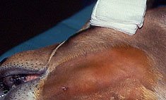
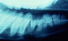
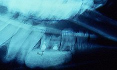
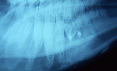
X-rays of the mandible of a Black Faced Impala


The patient
 
Close up View of the left mandibular swelling
 
Pre-operative Radiograph of the abscessed left mandibular 4th pre-molar showing loss of cortical bone along the lower boarder of the mandible and developing cementoma-like enlargements surrounding the root tip apexes.
 
Immediate Post-operative radiograph of the same tooth showing the amputated root tips; opaque retro-filled pulp canal spaces; and synthetic bone grafting material filling the marrow space.
 
A 6-month Post-operative View of the same area showing resolution of the bony defect with osseous integration of the synthetic grafting material providing re-establishment of a solid lower boarder to the mandible with a couple of small calcific nodules in.


For translation information and instructions, please CLICK HERE.

http://www.colyerinstitute.org
Website Content Copyright © 2009, The Colyer Institute
For copyright details on specific photos please
contact us by e-mail at info@colyerinstitute.org 

This web site is best viewed with a browser setting of 800 x 600 or greater.  If you use a smaller screen size and would like to view the content of this site using an alternate interface, please click here.