X-rays of the mandible of a Black Faced Impala
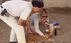
The patient
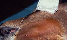
Close up View of the left mandibular swelling
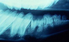
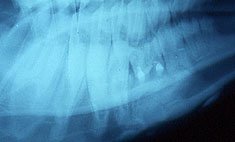
Pre-operative Radiograph of the abscessed left mandibular 4th pre-molar showing loss of cortical bone along the lower boarder of the mandible and developing cementoma-like enlargements surrounding the root tip apexes.
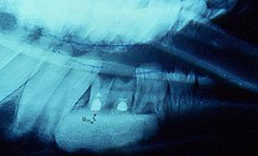
Immediate Post-operative radiograph of the same tooth showing the amputated root tips; opaque retro-filled pulp canal spaces; and synthetic bone grafting material filling the marrow space.
A 6-month Post-operative View of the same area showing resolution of the bony defect with osseous integration of the synthetic grafting material providing re-establishment of a solid lower boarder to the mandible with a couple of small calcific nodules in.