Oral Exams & Endodontic Treatment of 2 Black Footed Ferrets
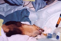
Ferret stabilized on inhalation anesthesia on a warm air pad
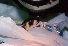
X-ray positioned for maxillary anterior dentition
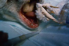
Excessive abrasive wear of the dentition in the female
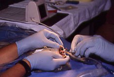
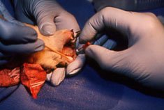
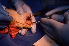
Utilizing an Electronic Apex Locating Device to determine the working length of the pulp canal of the upper right canine tooth.
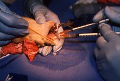
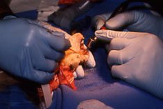
Enlarging, shaping and cleaning the pulp canal with a standard #25 H Endodontic instrument.
Flushing the canal with solutions of hydrogen peroxide and sodium hypochlorite
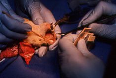
Drying the clean, disinfected canal with an endodontic paper point
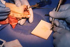
Utilizing a lentulo-spiral drill in a slow speed hand piece to fill the fill the canal with endodontic sealer paste.
Placing a fitted gutta-percha endodontic point to insure complete filling of the pulp canal space.
Final contouring of the composite resin restoration of the clinical crown of the tooth

Preparing to x-ray
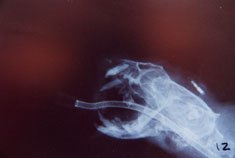
Final x-ray confirming endodontic treatment of the canine tooth