|
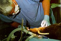
Restoration of a Sub-Gingival Tusk Fracture on a Male Babirusa |
||
 |
 |
|
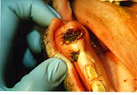
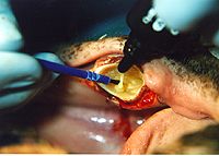
| The Initial Examination of the Fracture Site with View of this Species Uniquely Positioned Maxillary Tusk. | What’s left of the Sub-Gingivally Fractured Mandibular Tusk. | |
|
|
||
 |
 |
|
| Removing the Hyper-plastic Gingival Tissue to Find the Remaining Tooth Structure. | View of Secondary Dentine Bridge Protecting Underlying Vital Pulp Tissue. | |
|
|
||
 |
 |
|
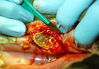
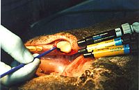
| The Process of Sealing, Protecting and Bonding to the Remaining Etched Dentin. | It Sometimes Requires a Bulk of material to Restore a Large Tooth. | |
|
|
||
 |
 |
|
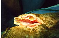
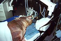
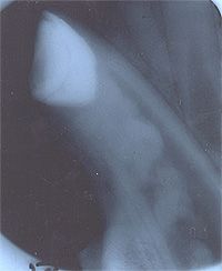
| The Restored Tooth is X-rayed to Document the Case for the Patient’s Medical Record. | Radiograph clearly Demonstrates Restoration, Secondary Dentin Bridge, Developing Pulp Stones, and Vital Pulp Tissue. | |
|
|
||
|
For translation information and instructions, please CLICK HERE. http://www.colyerinstitute.org
This web site is best viewed with a browser setting of 800 x 600 or greater. If you use a smaller screen size and would like to view the content of this site using an alternate interface, please click here. |
||