|
Oral Digital Radiology in an Indian Rhino |
||
|
|
|
|
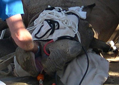
| View of palpation exam of Rhino’s right mandible. |
View of Digital Camera capturing
intra-oral view of healed fracture site to be printed for Clinical Record. |
|
|
|
||
|
|
|
|
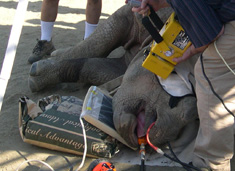
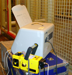
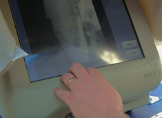
| View of Portable X-ray head projecting image of Rhino’s right mandible onto special Digital X-ray Plate attached by computer cable to CPU for projection onto computer’s monitor. | View of Portable X-ray Machine sitting next to Portable computer CPU/Monitor. | |
|
|
||
|
|
|
|
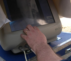
| View of digital X-ray monitor shaded to protect from direct sun light. | Closer view of image on computer screen. | |
|
|
||
|
|
||
|
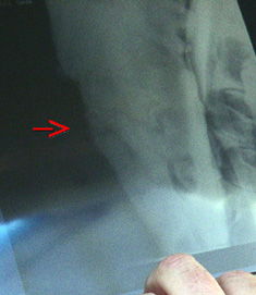
Close-up, enhanced view of red arrow
at healed mandibular fracture site on computer screen. |
||
|
|
||
|
|
||
|
For translation information and instructions, please CLICK HERE. http://www.colyerinstitute.org
This web site is best viewed with a browser setting of 800 x 600 or greater. If you use a smaller screen size and would like to view the content of this site using an alternate interface, please click here. |
||