|
Root Caries related PeriApical Abscess in a Cape Hunting Dog |
||
|
|
|
|
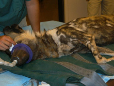
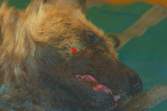
| View of dog during anesthesia induction. | Red Pointer indicates location of recurrent fistulating abscess below right eye. | |
|
|
||
|
|
|
|
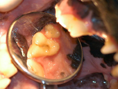
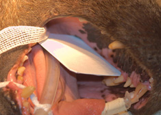
| Intra-oral view of maxillary right 1st molar. Notice this tooth presents a near perfect clinical crown without any sign of caries or fracture. | View of pre-packaged dental film in position within the oral cavity ready for exposure by x-ray. | |
|
|
||
|
|
|
|
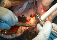
| X-ray image of molar tooth reveals significant apical abscessation involving both mesial and distal tooth roots. | Creating a “surgical moat” around tooth underneath a retracted full thickness muco-gingival soft tissue flap in order to facilitate removal of the tooth with minimal bony trauma using a high speed dental hand-piece with a cross cut fissure bur. | |
|
|
||
|
|
|
|
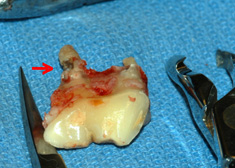
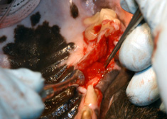
| View of freshly extracted molar tooth. Notice root caries at red arrow, confirming the fact that the apical abscess involving this tooth would NOT be resolvable with endodontic therapy alone. | View of extraction site while removing mass of infected soft tissue surrounding mesial root. Note: All of this infected tissue MUST be removed to fresh, clean bone in order to insure proper healing of the extraction site. | |
|
|
||
|
|
||
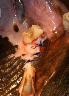
| View of extraction site sutured closed to protect Bioglass, particulate bone graft materal within. | ||
|
|
||
|
|
||
|
For translation information and instructions, please CLICK HERE. http://www.colyerinstitute.org
This web site is best viewed with a browser setting of 800 x 600 or greater. If you use a smaller screen size and would like to view the content of this site using an alternate interface, please click here. |
||