 |
|
 |
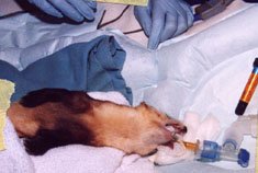
Ferret
stabilized on inhalation anesthesia on a warm air pad
|
|
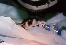
X-ray
positioned for maxillary anterior dentition
|
 |
|
 |
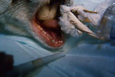
Excessive
abrasive wear of the dentition in the female
|
|
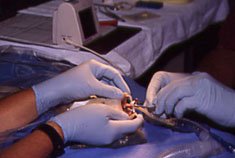
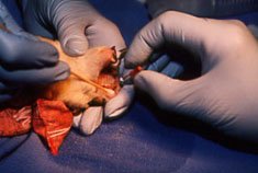
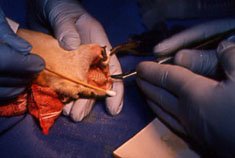
Utilizing
an Electronic Apex Locating Device to determine the working
length of the pulp canal of the upper right canine tooth.
|
 |
|
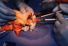 |
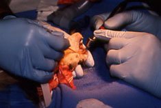
Enlarging,
shaping and cleaning the pulp canal with a standard #25 H
Endodontic instrument.
|
|
Flushing
the canal with solutions of hydrogen peroxide and sodium
hypochlorite
|
 |
|
 |
Drying
the clean, disinfected canal with an endodontic paper point
|
|
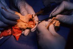
Utilizing
a lentulo-spiral drill in a slow speed hand piece to fill the
fill the canal with endodontic sealer paste.
|
 |
|
 |
Placing
a fitted gutta-percha endodontic point to insure complete
filling of the pulp canal space.
|
|
Final
contouring of the composite resin restoration of the clinical
crown of the tooth
|
 |
|
 |
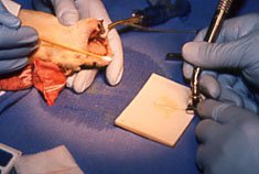
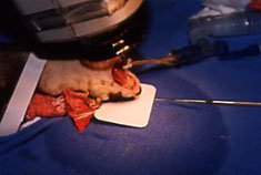
Preparing
to x-ray
|
|
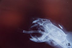
Final
x-ray confirming endodontic treatment of the canine tooth
|